
Figure 1.
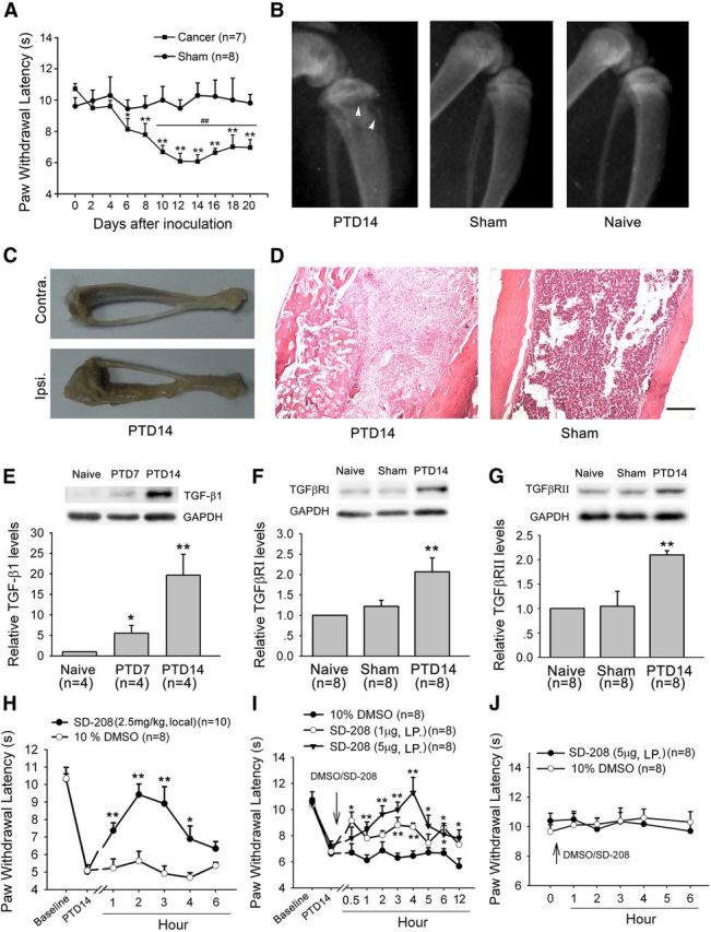
Involvement of bone-derived TGF-β1 and its receptors TGFβRI and TGFβRII in bone cancer pain. A, Intratibia inoculation with Walker 256 mammary gland carcinoma cells induces significant thermal hyperalgesia in the hindpaw ipsilateral to the tumor-bearing limb. *p < 0.05 versus basal (before tumor inoculation). **p < 0.01 versus basal (before tumor inoculation). ##p < 0.01 versus sham control. B, Radiographs represent robust radiolucent lesions (arrowhead) of the tibia on PTD 14. C, Photograph of a rat tibia on PTD 14 with obvious tumor growth in the ipsilateral but not contralateral tibia. D, Histopathological sections (hematoxylin and eosin stain) show that the bone marrow was largely replaced by invading tumor cells with medullary bone loss and tibial bone destruction on PTD 14. Scale bar, 500 μm. E, Western blot analysis reveals a progressive increase in the level of TGF-β1 in the affected bone after tumor inoculation. F, G, Western blot analysis reveals a significant increase in the level of TGFβRI and TGFβRII in L3–L5 DRGs ipsilateral to the tumor-bearing bone on PTD 14. *p < 0.05 versus naive. **p < 0.01 versus naive. H, Local injection (around tumor bone) of SD-208 (2.5 mg/kg) attenuated bone cancer-induced thermal hyperalgesia on PTD 14. I, Lumbar puncture injection of SD-208 (1 and 5 μg) significantly reduces bone cancer-induced thermal hyperalgesia on PTD 14. J, Lumbar puncture injection of SD-208 does not alter the paw withdrawal response to radiant heat stimulation in naive rats. *p < 0.05 versus vehicle (DMSO). **p < 0.01 versus vehicle (DMSO).