
Figure 5.
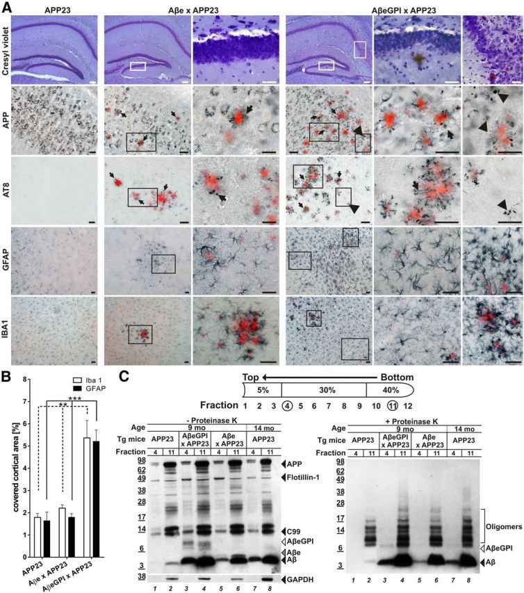
AβeGPI exacerbates amyloid-associated toxicity and promotes localization of aggregated Aβ species in DRM. A, Degenerative processes in double tg Aβe × APP23 and AβeGPI × APP23 mice. Hippocampal brain sections of 9-month-old tg mice stained with cresyl violet, anti-APP, anti-Tau (AT8), GFAP, and Iba1, respectively. All sections were additionally stained with Congo red. Higher magnifications for the double tg mice are shown in the right columns. In double tg AβeGPI × APP23 mice, displaced neurons and neuron loss are found at the sites of plaque deposition in neuronal loss at the stratum granulosum of the dentate gyrus and the CA1 stratum pyramidale of the hippocampus. Both double tg mice show dystrophic APP-positive synaptic boutons (arrows) and AT8-positive neuritic structures (arrows) in proximity to congophilic plaques. In addition, in the AβeGPI × APP23 mice APP-positive and AT8-positive dystrophic structures are found in vicinity of amorphous and Congo red-negative Aβ deposits (arrowheads). Both double tg lines display activations of astrocytes and microglia. However, in the AβeGPI × APP23 mice additional activated glia cell are found that are not restricted to the direct proximity of the congophilic deposits. Scale bar, 40 μm. B, Morphometric analysis revealed a significant increase of the area covered by Iba1 and GFAP staining in double tg AβeGPI × APP23 mice compared with Aβe × APP23 and APP23 mice (n = 5 for APP23, n = 7 for Aβe × APP23, n = 10 for AβeGPI × APP23; **p < 0.01, ***p < 0.001, one-way ANOVA Bonferroni's post hoc test for multiple comparisons). C, Left DRM extraction from brain homogenate treated with cold Triton X-100, followed by OptiPrep step density gradient. DRM proteins (fraction 4) and total protein load (fraction 11) were compared in 9-month-old tg APP23, double tg AβeGPI × APP23, Aβe × APP23, and 14-month-old tg APP23 mice by Western blot probed with 6E10. Aβ is present in all DRM fractions except for the 9-month-old APP23 predepositing mice (lanes 1 and 2). Flotillin-1 marker for DRMs; GAPDH marker for cytoplasmic protein. Right, Fractions from left treated with 100 μg/ml proteinase K (+PK) for 30 min and analyzed by Western blot probed with Aβ antibody 6E10. DRM-associated Aβ of respective tg mice (lanes 3, 5, and 7) displayed PK resistance.