
Figure 1.
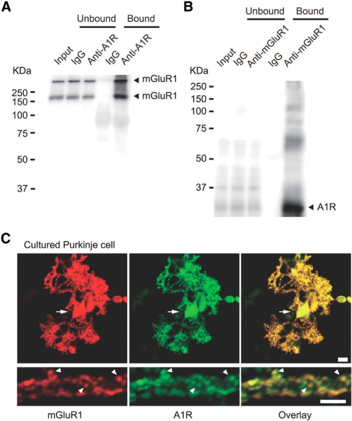
Endogenous interaction between A1R and mGluR1 in cerebella and colocalization of A1R and mGluR1 in cultured Purkinje cells. A, Coimmunoprecipitation of mGluR1 with A1R in the lysate of the crude synaptosome fractions derived from mouse cerebella. Monomeric and dimeric bands were detected by immunoblotting with anti-mGluR1 antibody in the anti-A1R immunoprecipitate. B, A1R was detected by immunoblotting with the anti-A1R antibody in the anti-mGluR1 immunoprecipitate. C, Double-immunofluorescence staining of a cultured Purkinje cell (16-d-old in vitro) with anti-mGluR1 (red) and anti-A1R (green) antibodies. Arrows and arrowheads, soma and dendritic spines with colocalized signals, respectively. Scale bars, 10 μm.