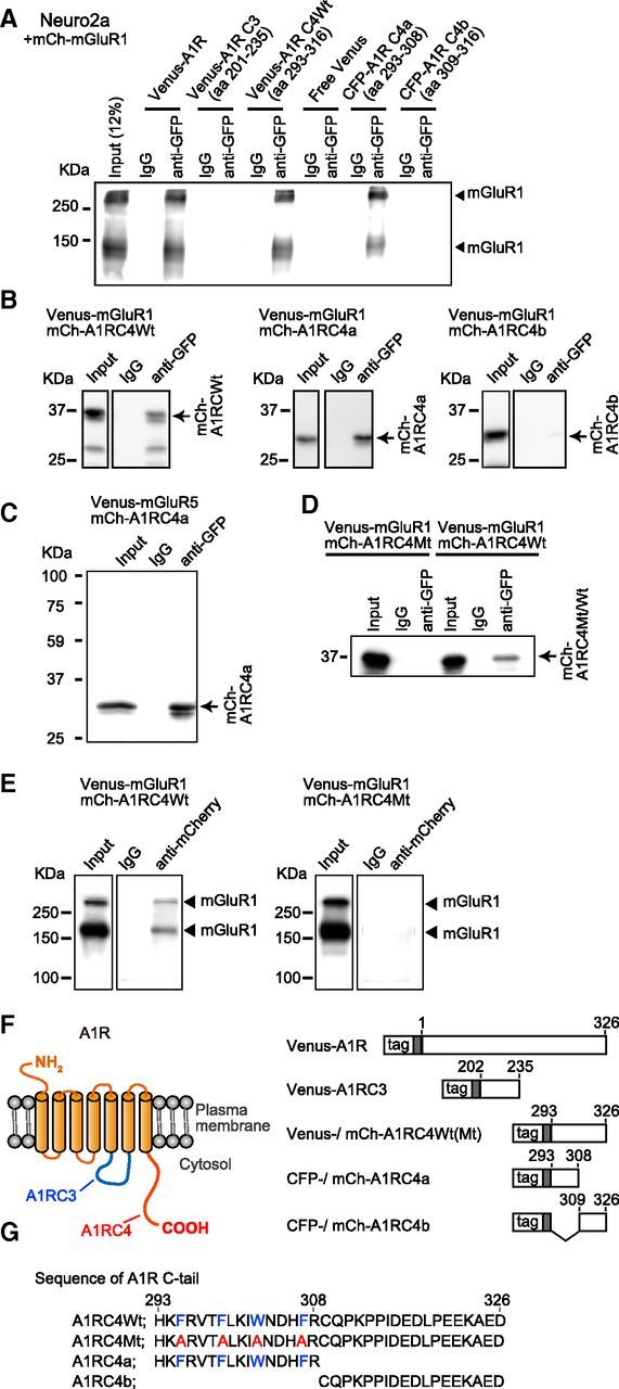
Figure 2.

Complex formation of A1R and mGluR1 in Neuro2a cells. A, Coimmunoprecipitation of mCherry-mGluR1 with Venus- or CFP-fused full-length A1R or A1R C-terminal tail. Lysates from Neuro2a cells expressing Venus- or CFP-fused full-length or cytoplasmic domains of A1R and mCherry-mGluR1 were immunoprecipitated with the rabbit anti-GFP antibody. The precipitated fractions were immunoblotted with the mouse anti-mGluR1 antibody. B, Coimmunoprecipitation of mCherry-fused full-length A1R and A1R C-terminal tail with Venus-mGluR1. Neuro2a cells were transfected with Venus-mGluR1 and either mCherry-fused A1R C-tail (mCh-A1RC4Wt, aa 293–316), mCherry-fused A1R C-tail membrane-proximal region (mCh-A1RC4a, aa 293–308), or mCherry-fused the A1R C-tail membrane-distal region (mCh-A1RC4b, aa 309–316). Crude cell lysate (Input) and the immunoprecipitates with the anti-GFP or control IgGs were immunoblotted with the anti-mCherry antibody. C, Immunoprecipitation with the anti-GFP antibody from the extracts of Neuro2a cells expressing Venus-mGluR1 and either mCh-A1RC4Wt or a mCherry-fused mutant A1R C-tail (mCh-A1RC4Mt). In mCh-A1RC4Mt, F295, F299, W303, and F307 were replaced with alanines. Crude cell lysate (Input) and the immunoprecipitates with the anti-GFP or control IgGs were immunoblotted with the anti-mCherry antibody. Arrow, mCherry-fused A1RC4Wt, A1RC4a, or A1RC4b. D, Immunoprecipitation with the anti-mCherry antibody from the extracts of Neuro2a cells expressing Venus-mGluR1 and either mCh-A1RC4Wt or mCh-A1RC4Mt. Crude cell lysate (Input) and the immunoprecipitates with the anti-mCherry or control IgGs were immunoblotted with the anti-GFP antibody. Filled triangles, mGluR1 monomer and dimer. E, Schematic structure of A1R and its cytoplasmic domains used for the coimmunoprecipitation studies. F, Amino acid sequence of the A1R C-terminal tail and the alanine mutant.
