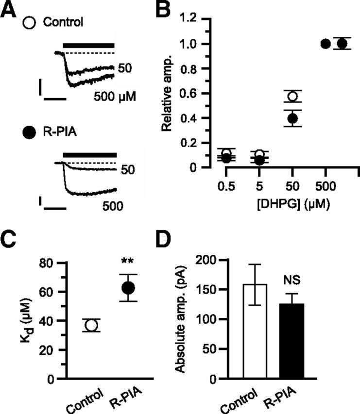
Figure 6.

A1R activation decreases mGluR1's ligand sensitivity. A–D, DHPG-induced inward currents in the absence (Control, n = 10) or presence of R-PIA (50 nm, R-PIA, n = 10). A, Each set of superimposed traces indicates sample inward current evoked by saturating (500 μm) and unsaturating (50 μm) doses of DHPG in a Purkinje cell. Holding potential, −70 mV. Scale bars, 10 s and 50 pA. B, Mean relative peak amplitude of the inward currents as a function of DHPG dose. The amplitude was normalized to the value with 500 μm DHPG for each cell. C, Mean apparent Kd. D, Mean absolute peak amplitudes of inward currents evoked by the saturating dose of DHPG. **p < 0.01; NS, p > 0.05, unpaired t test. Error bars, ±SEM.
