Figure 7.

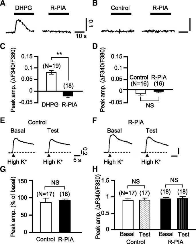
A1R activation does not induce an elevation of [Ca2+]i in Purkinje cells. A, [Ca2+]i responses to R-PIA (50 nm, 30 s) were measured in the cells that displayed Ca2+ release from the intracellular stores in response to DHPG (50 μm, 20 s). Thick bars, timing of drug application; vertical scale bars, changes in F340/F380. B, [Ca2+]i responses to R-PIA were measured in the cells that had been exposed only to the vehicle, not to DHPG. C, D, Mean peak amplitudes of the [Ca2+]i responses to the labeled drugs measured as shown in A and B. R-PIA did not increase the cytoplasmic Ca2+ level, indicating that A1R activation with the submicromolar level of the agonist does not facilitate constitutive Ca2+ release. E, F, Each set of traces indicates sample depolarization-evoked [Ca2+]i rises obtained from a cell before (Basal) and after (Test) a 12 min local application of the normal (Control) or R-PIA (50 nm)-containing saline (R-PIA). To depolarize the cells, high-K+ (75 mm)-containing saline was applied locally for 1 s (arrowheads). Vertical scale bars, changes in F340/F380. G, Mean peak amplitudes of the [Ca2+]i rises after an application of the normal saline or R-PIA measured as shown in E and F. Amplitude is expressed as the percentage of the basal level for each cell. H, Mean resting [Ca2+]i before and after an application of the normal saline or R-PIA. **p < 0.01; NS, p > 0.05, paired t test. Error bars, ±SEM.
