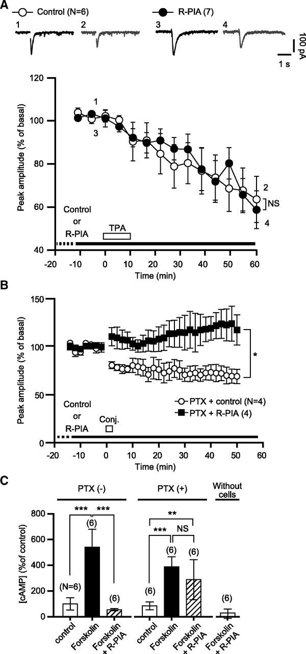
Figure 8.

A1R-mediated blockade of glu-LTD is not due to modulation of the PKC or PKA cascade. A, Each pair of traces indicates sample Iglu of a cell before and after a bath application of TPA (200 nm, 10 min) in the absence (Control) or continuous presence of R-PIA (50 nm). Each plot indicates the time course of the mean peak amplitude of Iglu. White bar, timing of TPA application; dots and error bars, mean ± SEM of the data of every 5 min period. B, PTX, a Gi/o-protein inhibitor, does not abolish R-PIA-induced glu-LTD blockade. Each plot indicates the time course of the mean peak amplitude of Iglu of a PTX (500 ng/ml, over 16 h)-pretreated cell before and after the conjunctive depolarization/glutamate stimuli in the absence (Control) or continuous presence (R-PIA) of R-PIA (100 nm). Dots and error bars, mean ± SEM of the data for every 2 min period. C, Comparison of the effects of the labeled test agents on the cAMP production of the HEK293 cells stably expressing A1R and mGluR1 by LANCE cAMP competitive immunoassay. The assay was performed after a 30 min test agent application. PTX(-), cells-without a PTX pretreatment; PTX(+), PTX (500 ng/ml, > 12 h)-pretreated cells. Error bars, ±SD. Without cells, mock-up assay with cell-free buffers.
