Figure 2.

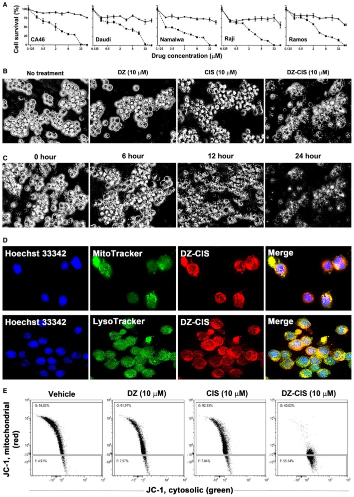
Heptamethine carbocyanine fluorescent dye–cisplatin conjugate (DZ‐CIS) cytotoxicity. (A) Dose‐effect curve. All Burkitt lymphoma cell lines were insensitive to CIS (circles on solid line) but dose‐dependently sensitive to DZ‐ CIS (squares on dotted line). (B) DZ‐CIS‐induced death of Namalwa cells 24 hours after treatment. Black and white images of trypan blue–stained cells (magnification ×100). (C) DZ‐CIS (10 μM) caused marked lymphoma cell death with morphological changes starting 6 hours into the treatment. Cell death became conspicuous around 12 hours (trypan blue stain, magnification ×100). (D) DZ‐CIS preferentially accumulates to mitochondria and lysosomes, demonstrated by colocalization (magnification ×600) of the NIR signal with MitoTracker (upper row) and LysoTracker (lower row). (E) Flow cytometry indicates that 6‐hour treatment with DZ‐CIS disrupts mitochondrial membrane integrity. JC‐1 in the cytosol lost its red fluorescence as monomeric JC‐1 (which emits green fluorescence).
