Figure 3.

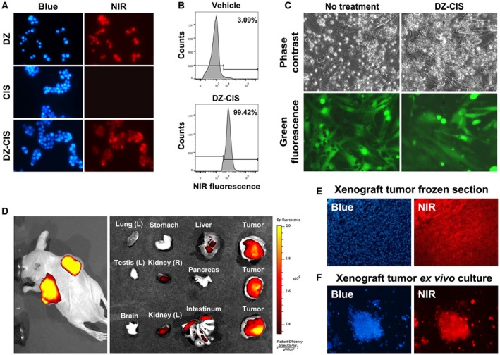
The heptamethine carbocyanine fluorescent dye–cisplatin conjugate (DZ‐CIS) acts specifically on Burkitt lymphoma cells. (A) Near‐infrared (NIR) microscopy of Namalwa cells after treatment with 4 μM DZ‐CIS for 15 minutes. DZ‐CIS accumulated in all lymphoma cells. Blue fluorescence of the nuclei was from Hoechst 33342 co‐stain (200×). (B) Namalwa cells treated with 4 μM DZ‐CIS for 15 minutes were assayed for rapid uptake of the fluorescent dye–drug conjugate in live cells using flow cytometry. (C) Images of lymphoma and stromal cell coculture show that DZ‐CIS preferentially kills lymphoma but not stromal cells. In coculture with GFP‐tagged CCD16Lu normal human lung mesenchymal stromal cells, 24‐hour DZ‐CIS treatment preferentially killed Burkitt lymphoma cells (Namalwa), while stromal cells survived and displayed healthy morphology (magnification ×200). DZ‐CIS–induced death of lymphoma cells in coculture was confirmed with trypan blue stain. (D) Tumor‐specific uptake and retention of DZ‐CIS in vivo were evaluated with NCrnu/nu mice bearing Namalwa xenograft tumors. After treatment with DZ‐CIS for 2 weeks, mice were subjected to whole body NIR imaging (left). Necropsy tumor samples together with host organs were subjected to ex vivo NIR imaging (right). (E) Frozen sections of xenograft tumors were stained with 4’,6‐diamidino‐2‐phenyindole dihydrochloride (DAPI) and examined for retention of DZ‐CIS by NIR microscopy (magnification ×100). (F) Xenograft tumors diced in ex vivo culture were stained with DAPI (magnification ×100). Most tumor cells still carried DZ‐CIS signals after 3 days in the culture.
