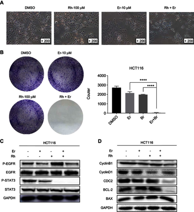
Figure 6.
Rhein and erlotinib efficiently synergistically suppresses phosphorylation of STAT3 and EGFR. (A) Morphological changes of HCT116 colon cancer cells observed by light microscopy after drug treatment. (B) Colony formation assay of HCT116 cells. (C) HCT116 cells were treated, in combination or separately, with erlotinib (Er; 10 μM) and/or rhein (Rh; 50 μM) for 24 h. P-STAT3, P-EGFR, STAT3 and EGFR were detected by Western blot analysis. GAPDH was used as the control protein. (D) HCT116 cells were treated, in combination or separately, with erlotinib (10 μM) and/or rhein (50 μM) for 24 h. CDC2, CyclinB1,CyclinD1,BCL2 and BAX were detected by Western blot analysis, GAPDH was used as the control protein, ****P<0.0001.

