
Figure 1.
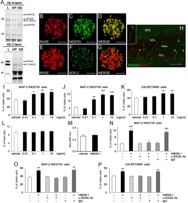
RAGE, expressed by adult hippocampal NPCs, mediates proneurogenic effects elicited by HMGB-1. A, Representative immunoblots confirming the presence of different RAGE isoforms in extracts from undifferentiated neural progenitors grown in NS, HP, or lung (L) by using two antibodies (Ab) recognizing different N- and C-terminal epitopes. Bands corresponding to RAGE-specific isoforms are marked. B–G, Representative fluorescent microscopy images of NS immunolabeled with antibodies against RAGE (B, E, red), nestin (C, green), and Sox-2 (F, green). The merged images showed that RAGE immunoreactivity colocalized with nestin (D) and Sox-2 (G). Magnification, 600×. Scale bar, 56.2 μm. H, Representative confocal microscopy image of the adult mouse DG immunostained with antibodies against RAGE (green) and Sox-2 (red). Some Sox-2+ NPCs (indicated by arrowheads) were also positive for RAGE. MOL, Molecular layer. Magnification, 400×. Scale bar, 70 μm. Inset, Higher magnification of a Sox-2+/RAGE+ cell. I–K, Under differentiating conditions, 24 h treatment of adult hippocampal NPCs with 0.1–10 ng/ml HMGB-1 significantly increased, in a concentration-dependent manner, the percentage of MAP-2+/nestin+ (I), MAP-2+/nestin− (J), and CR+ (K) cells compared with vehicle-treated cells. L, M, No significant difference was observed between vehicle- or HMGB-1-treated cultures in the percentage of apoptotic nuclei (L) and in LDH activity in media (M) of differentiated cell cultures. N–P, A neutralizing anti-RAGE antibody (α-RAGE, 20 μg/ml), but not preimmune IgG serum (IgG), abolished the proneurogenic effects of 1 ng/ml HMGB-1 on MAP-2+/nestin− (N), MAP-2+/nestin+ (O), and CR+ (P) cells. I–P, Data represent the mean ± SEM of n = 3 experiments, run in triplicates. *p < 0.05, **p < 0.01, ***p < 0.001 versus vehicle (Student's t test).