
Figure 3.
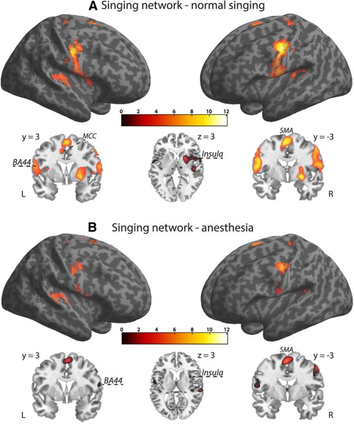
Brain activation of overt singing including all participants. A, Normal singing. B, Singing with anesthetized vocal-fold mucosa. During singing under normal feedback conditions, we found activation in regions that constitute the “singing network.” The same basic network was also revealed during singing with anesthetized vocal-fold mucosa, but at reduced activation strength. Activation maps were superimposed using the cortex_20484.surf template in SPM8, thresholded at p = 0.07 (FWE corrected) for visual display (extent threshold, 10 voxels) and the MRIcron software. Axial and coronal brain slices through the rendered volumes (shown below the corresponding images) illustrate frontomedial (BA44), premotor (SMA), subcortical (globus pallidus, putamen), and limbic sensorimotor areas [anterior insula and middle cingulate cortex (MCC)] during normal and anesthesia singing respectively.