Figure 7.
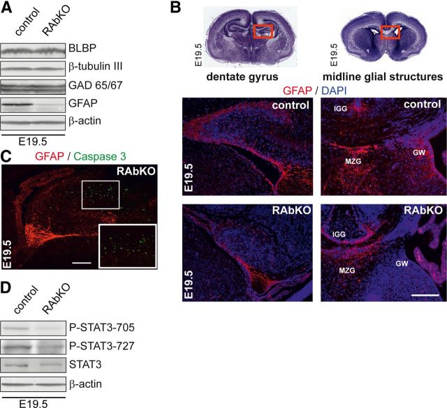
Astrocytic differentiation is impaired in RAbKO brains in vivo. A, Western blot analysis of E19.5 brain lysates probed for indicated markers of neural development. B, Top, Cresyl violet stainings of brain sections from E19.5 mice. Red boxes represent regions shown below: sections of dentate gyrus and midline glial structure regions stained with antibody to GFAP (red) and DAPI (blue). IGG, Indusium griseum glia; MZG, midline zipper glia; GW, glial wedge. C, Analysis of apoptotic cells in E19.5 hippocampus of RAbKO mice stained for GFAP and Caspase 3. Apoptotic cells are not GFAP-positive. The white frame represents the magnified inset. D, Western blot analysis of E19.5 brain lysates probed for indicated proteins. Scale bars: B, C, 200 μm.

