Figure 1.
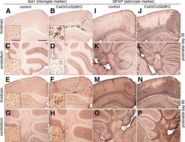
Activated microglia and reactive astroglia in the CNS of Cx43/Cx32dKOmGFAP mice. Immunohistochemical analysis on 25 μm brain slices from 30- and 90-d-old mice were stained for Iba1, a microglia marker. Control mice showed normal distribution of ramified microglial cells in all time points and regions tested (A, C, E, G). In Cx43/Cx32dKOmGFAP mice, a strong microglial activation along the corpus callosum and in the cingulum was detected at P30 (B), whereas the cerebellum was not affected (D). With advancing age, the intensity of the microglial activation in the cingulum decreased (F), but on P90, microglial activation occurred in the cerebellar white matter (H). Immunohistochemical analysis of GFAP on brain slices of 30- and 90-d-old controls showed the typical distribution of GFAP-positive cells in cortex and cerebellum (I, K, M, O). GFAP is only weakly expressed in the corpus callosum and in some astrocytes surrounding blood vessels in the cortex, whereas it is prominent in the hippocampus. In contrast, GFAP expression was highly increased in corpus callosum and especially cortex of Cx43/Cx32dKO mice on P30 (J). There were no differences between control (K) and dKO (L) mice in cerebellar GFAP expression. During maturation, astrogliosis in the cortex of Cx43/Cx32dKO mice increased. Nearly the entire cortical area was covered by strongly GFAP-positive cells on P90 (N), and astrogliosis was additionally detected in the cerebellum of dKO mice on P90 (P). Scale bars: 500 μm; insets, 100 μm.

