Figure 3.
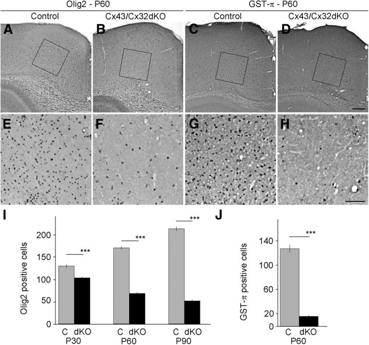
Number of oligodendrocytes is decreased in cingulum of Cx43/Cx32dKOmGFAP mice. Olig2-positive cells have been stained by immunohistochemistry in brain slices of 60-d-old mice. Normal number and distribution of Olig2-positive cells can be seen in brain slices of WT animals (A), whereas there were less Olig2-positive cells in the cingulum of Cx43/Cx32dKO mice (B). For better visualization, we provide higher magnifications of WT (E) and Cx43/Cx32dKO (F) mice. To quantify the number of myelinating oligodendrocytes, immunohistochemical staining against GST-π was performed on brain slices of 60-d-old mice. By comparing WT mice (C, G) with Cx43/Cx32dKO mice (D, H), a dramatic decrease of myelinating cells in the dKO animals becomes obvious. The number of Olig2-positive cells per 202,500 μm2 has been quantified for mice aged P30, P60, and P90 (I), whereas the number of GST-π-positive cells has only been quantified on P60 (J). Scale bars: A–D, 200 μm; E–H, 100 μm. ***p < 0.001.

