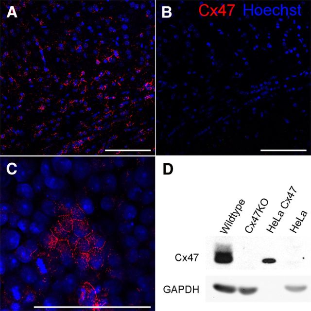
Figure 4.
Validation of newly generated Cx47 antibodies by immunofluorescence and immunoblot analysis. Immunofluorescence analysis using our newly generated rabbit polyclonal Cx47 antibodies in the cingulum of WT (A) and Cx47-deficient (B) mouse brain slices. Cx47 signals are shown in red, and nuclear staining by Hoechst33258 is shown in blue. In addition, anti-Cx47 reactivity was confirmed using Cx47-expressing HeLa cells (C). Immunoblot analyses (D) using 50 μg of cerebellar tissue lysate show strong Cx47 expression in WT but no Cx47 expression in Cx47KO cerebellum. HeLa WT and HeLa–Cx47 cells were used as controls, whereas GAPDH was used for standardization of immunoblots. Scale bars, 100 μm.

