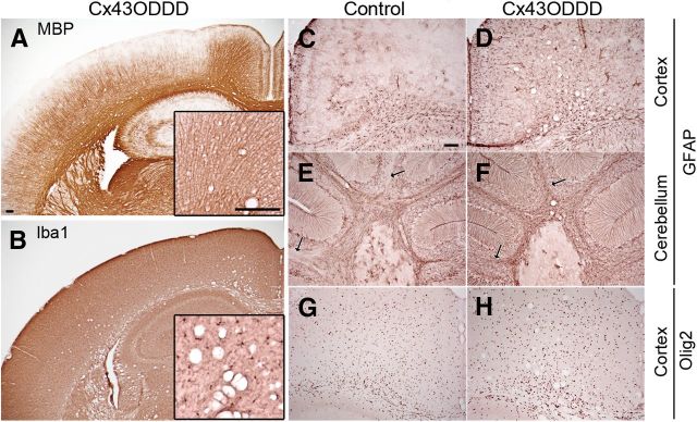
Figure 8.
Myelin, microglia, astrocytes and oligodendrocytes in Cx43ODDD mice. Immunostainings on brain slices of 50-d-old mice show normal myelination (A) and non-activated microglial cells (B). Strong astrogliosis is visible in cortex and cerebellum (arrows) of Cx43ODDD mice (D, F) compared with control mice (C, E). Normal distribution of Olig2-positive oligodendrocytes in cingulum and cortex of Cx43ODDD mice (H) compared with controls (G). Vacuoles are mostly found in white matter regions across the brain of Cx43ODDD mice (A, B, D, F, H). Scale bars, 100 μm.

