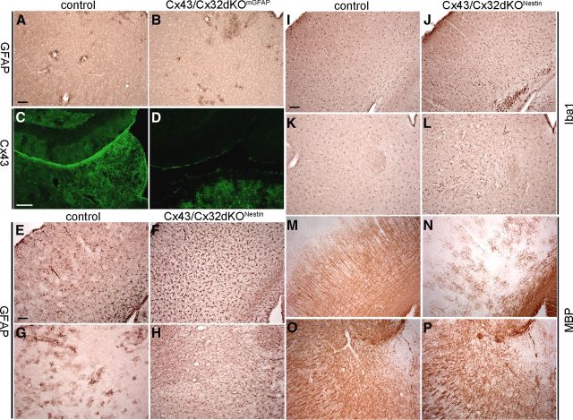
Figure 9.
Immunohistochemical analyses of Cx43/Cx32dKONestin mice and comparison with Cx43/Cx32dKOmGFAP mice. Immunohistochemical staining for GFAP on brain slices of 60-d-old Cx43/Cx32dKOmGFAP mice revealed normal distribution of GFAP-positive astrocytes in the thalamus (A, B). mGFAP–Cre-driven deletion of Cx43 is incomplete; immunostainings for Cx43 show several Cx43-positive puncta remaining in the thalamus of Cx43/Cx32dKOmGFAP mice (C, D). Analyses of the phenotypic alterations in Cx43/Cx32dKONestin mice were performed by immunohistochemical stainings for GFAP, Iba1, and MBP on brain slices of 60-d-old-mice. Control mice exhibit normal distribution of GFAP-positive astrocytes in the cortex (E) and thalamus (G), whereas Cx43/Cx32dKONestin mice suffer from strong astrogliosis in the cortex (F) and thalamus (H). This is in contrast to Cx43/Cx32dKOmGFAP mice, which show no astrogliosis in the thalamus. Control mice show normal morphology of microglial cells (I, K), but in Cx43/Cx32dKONestin mice, microglial cells are activated throughout myelinated areas (J, L). Myelin staining shows strong loss of fine myelinated fibers in the cortex of Cx43/Cx32dKONestin mice (N) compared with controls (M). In the thalamus, the myelin appears less dense in Cx43/Cx32dKONestin mice (P) compared with controls (O). Scale bars, 100 μm.

