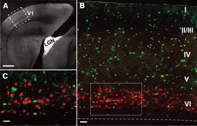
Figure 1.
Identification of neurons that project to the dorsal lateral geniculate nucleus (CG cells). A, The dorsal lateral geniculate nucleus (LGN) into which Alexa Fluor 555-conjugated cholera toxin subunit B had been injected is shown as a white area. The primary visual cortex is indicated by V1. An area boxed by dotted lines is shown in B as a magnified fluorescent image. Scale bar, 500 μm. B, GABAergic neurons are shown in green and CG cells are shown in red. Cortical layers are marked by Roman numbers in the right. Dashed line indicates the border between layer VI and WM. Scale bar, 60 μm. C, Magnified image of the area boxed by dotted lines in B. Scale bar, 20 μm.

