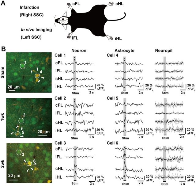
Figure 1.
Effect of unilateral SSC stroke on limb-stimulation-induced intracellular calcium concentration changes in neurons and astrocytes in contralateral SSC. A, Schematic drawing of experimental design. All strokes were made in right SSC, and images or samples were taken from left SSC. B, Representative TPLM images (left panels) and traces of changes in intracellular calcium level in numbered cells (right panels) in the mice of sham-operated (Sham), first week (5–7 d after stroke), and second week (8–12 d after stroke) after the stroke (1 week and 2 week groups, respectively). Each trace indicates the ΔF/F0% of calcium transients induced by limb stimulation. In the case of neuropils, responses from different neuropils (arrowheads) were shown in thin lines. Their average was shown in the thick line. The average of response from neuropils was not above the threshold for signal analysis.

