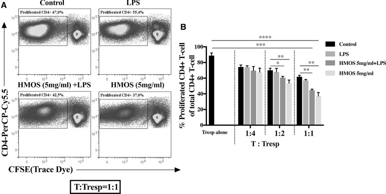
Figure 6.

Suppressive capacity of HMOS‐conditioned human moDCs on activated responder CD4+ T‐cell proliferation. (A) Representative plots of T‐responder cell (Tresp) co‐cultured with CD4+T‐cell primed by control DCs, LPS treated DCs, HMOS (5 mg/ml) +LPS treated DCs, or HMOS (5 mg/ml) treated DCs at the ratio of 1:1. (B) The degree of Tresp proliferation after co‐culturing with CD4+T‐cell primed by different DCs. CD3/CD28 activated CFSE‐labelled responder CD4+T‐cell was co‐cultured with CD4+T‐cell primed by different DCs at ratio 4:1, 2:1, and 1:1 for 5 days. Proliferation of FITC‐positive cells was analyzed by flow cytometry and suppressive functionality was determined by comparing the proliferated CD4‐PerCP‐Cy5.5 positive T cell. Results are presented as mean ± SEM, (3 independent experiments (1‐2 donors/experiment). *p < 0.05, **p < 0.01, ***p < 0.01, ****p < 0.001, paired Student's t‐test.
