
Figure 8.
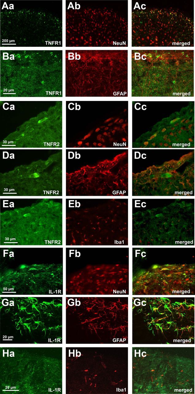
Receptors for the pro-inflammatory cytokines TNF-α and IL-1β are expressed by lamina I neurons and glial cells. Double immunostaining shows colocalization of TNFR1 (Aa,Ba, green) and TNFR2 (Ca,Da,Ea, green) and IL1R (Fa,Ga,Ha, green) with the neuronal marker NeuN (Ab,Cb,Fb, red), the astrocytic marker GFAP (Bb,Db,Gb, red), and the microglial marker Iba1 (Eb,Hb, red) in spinal cord lamina I. Overlays reveal that lamina I neurons express TNFR1, TNFR2, and IL-1R. TNFR1 and IL-1R are also expressed by GFAP-positive cells in the spinal dorsal horn. There is weak costaining for the individual receptors and the microglial marker Iba1.