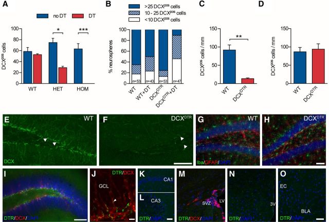
Figure 1.
Depletion of immature neurons in DCXDTR knock-in mice. A, DT ablated DCX+ cells in cultures of embryonic cortical neurons derived from heterozygous (HET; n = 3) and homozygous (HOM; n = 4) DCXDTR mice but not from WT controls (n = 2). B, Addition of DT to differentiated neurospheres ablated DCX+ cells. Note the dramatic reduction in the number of DCX+ cells encountered within neurospheres from DCXDTR mice after the addition of DT. C, In vivo DT administration resulted in depletion of DCX+ cells in the dentate gyrus of DCXDTR mice (n = 3) but not WT controls (n = 3). D, Recovery of DCX+ cell numbers in the dentate gyrus in DCXDTR mice was observed 28 d after the last DT injection (n = 4 per condition). E, F, Representative images showing depletion of DCX+ cells in the dentate gyrus of DCXDTR but not WT mice after DT treatment. White arrowheads indicate DCX+ cells. G, H, Representative images of the distribution of Iba1+ microglia and GFAP+ astrocytes in WT and DCXDTR mice after DT treatment. I, Photomicrograph showing staining for DTR (green) and DCX (red) in the dentate gyrus of DCXDTR mice (cell nuclei are counterstained blue with DAPI). J, Higher-power image showing punctate DTR staining (green) in close association with DCX+ cells (red) and their processes (white arrowhead). K–O, No detectable DTR staining was observed in other regions of the hippocampus (i.e., the CA1 and CA3) (K,L), the SVZ (M), hypothalamus (N), or amygdala (O). Scale bars: E, F, 100 μm; I, 70 μm; J, 10 μm; K–O, 50 μm. *p < 0.05. **p < 0.01. ***p < 0.001. GCL, Granule cell layer; LV, lateral ventricle; 3V, third ventricle; BLA, basolateral amygdala; EC, external capsule.

