
Figure 1.
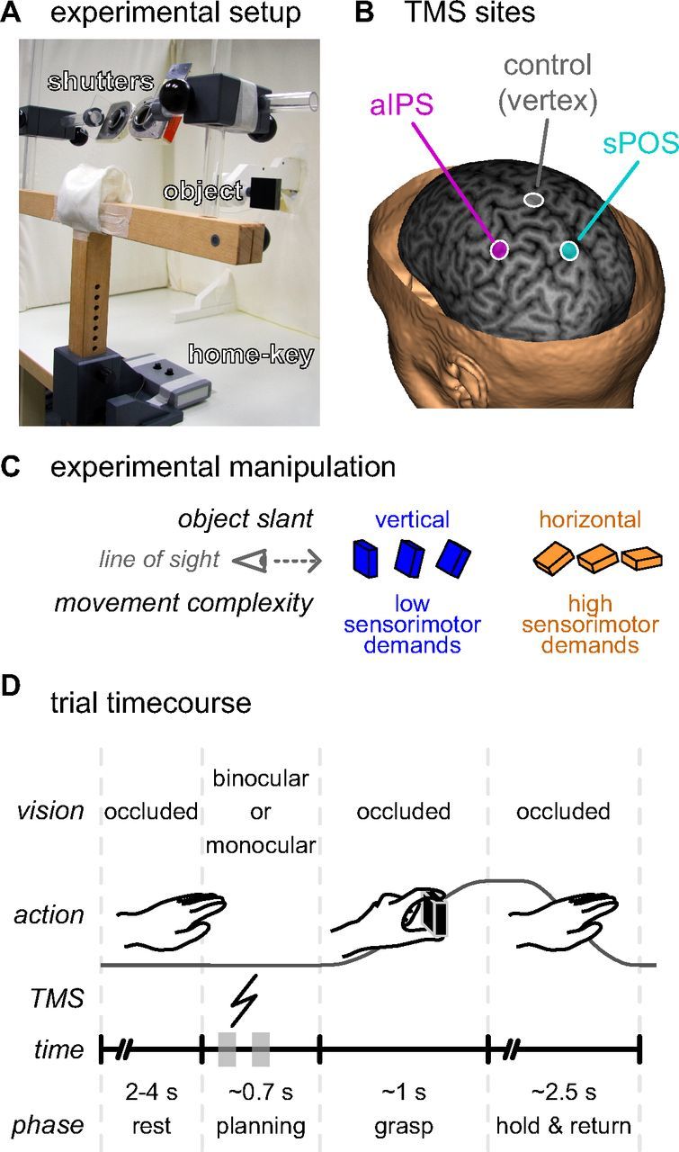
Experimental setup and task. A, The subject was seated at a table with his or her head resting in a chin-rest, the right hand on a home-key, and vision occluded by two shutters. These shutters could open individually to allow vision of the object to be grasped (a 6 × 6 × 2 cm black rectangular object), which was suspended in front of the subject. B, A TMS coil was positioned over the subject's head targeted at one of three sites: (1) the anterior part of the left intraparietal sulcus (magenta, aIPS), (2) the anterior bank of the superior part of the parieto-occipital sulcus in the left hemisphere (cyan, sPOS), and (3) the vertex of the head (gray) serving as a control site. TMS target sites are projected on a curvilinear reconstruction of the structural MRI of one exemplary subject, shown here at 6 mm in depth from the cortical surface. The location of the aIPS and sPOS sites were based on group averaged fMRI activation coordinates from a previous study (Verhagen et al., 2008). C, To vary the sensorimotor demands of the grasping task we manipulated the slant of the target object along the midsagittal plane: in seven slants from vertical to horizontal relative to the frontoparallel plane, in 15° steps. D, At the start of each trial, after a random interval (2–4 s) with occluded vision, either monocular or binocular vision of the target object was provided. The subject was instructed to prepare and perform a grasping movement, displacing the target object before returning to the starting position. During the planning phase, a single TMS pulse was delivered between either 100–200 ms or 300–400 ms after stimulus presentation (indicated on the trial time course with gray blocks). As soon as the subject released the home-key to grasp the object, the shutter glasses closed, preventing visual feedback of the movement.