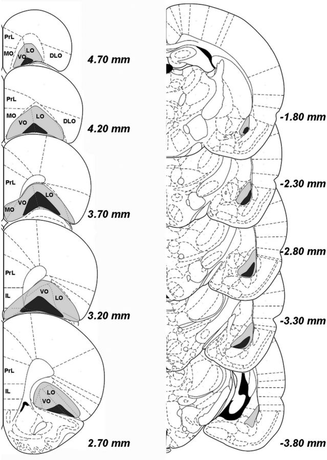
Figure 2.
Extent of lesion in the OFC or BLA. The extent of damage to the largest (light gray) and smallest (dark gray) regions of the OFC (left) and BLA (right), regardless of hemisphere, are depicted schematically on the right hemisphere. DLO indicates dorsolateral prefrontal cortex; IL, infralimbic cortex; LO, lateral orbitofrontal cortex; MO, medial orbitofrontal cortex; PrL, prelimbic cortex; VO, ventral orbitofrontal cortex.

