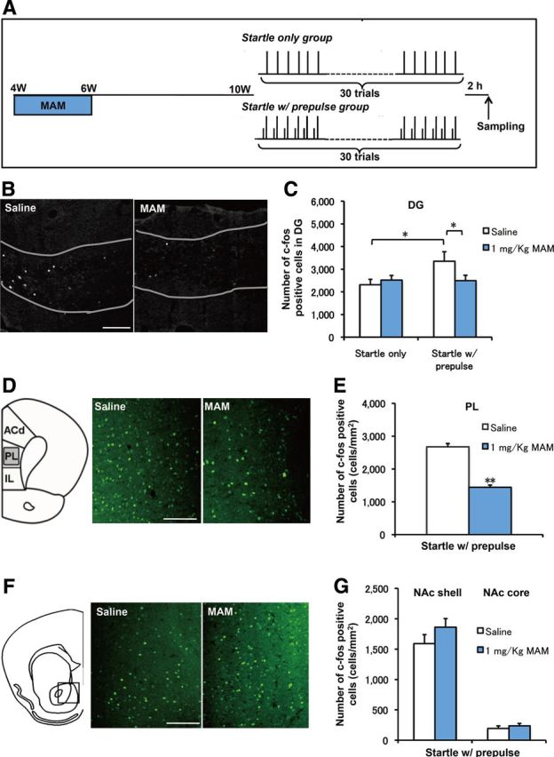
Figure 4.

PPI-induced hippocampal activation was blocked by MAM treatment. A, Experimental design of MAM treatment and the behavioral manipulation protocol. B, Representative c-fos staining in the DG. C, There was an increase in the number of c-fos+ cells in the startle-with-prepulse group compared with the startle-only group (F(1,14) = 4.62166, p = 0.04953 startle with prepulse plus saline vs startle only plus saline, n = 8 for each group), but this increase was blocked by MAM treatment during PW4–PW6 (F(1,16) = 4.54244, p = 0.4892 for startle with prepulse plus MAM vs startle with prepulse plus saline, n = 8 for startle with prepulse plus saline and n = 10 for startle with prepulse plus MAM). D, Representative c-fos staining in the PL cortex. E, MAM treatment decreased the number of c-fos+ cells in the PL cortex (F(1,18) = 89.19019, p < 0.001 for startle with prepulse plus MAM vs startle with prepulse plus saline, n = 10 for each group). E, Representative c-fos staining in the nucleus accumbence (NAc). F, There were no changes in c-fos+ cells in the NAc shell and NAc core between groups (NAc shell: F(1,20) = 1.79051, p = 0.19588 for MAM vs saline control, n = 11 for each group; NAc core: F(1,14) = 0.56137, p = 0.4611 for MAM vs saline n = 8 mice for each group). Scale bar, 200 μm. *p < 0.05; **p < 0.01.
