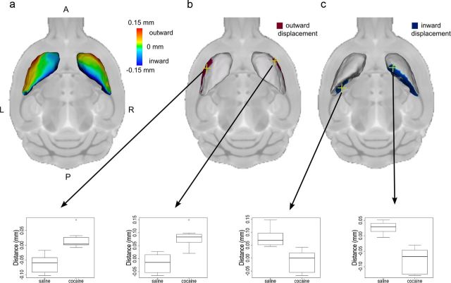
Figure 2.
Striatum shape was altered by cocaine exposure. Group differences in striatum surface position show that the lateral surface of the striatum was displaced outward (away from the center of the structure) and the medial surface of the striatum was displaced inward (toward the center of the structure) in mice exposed to cocaine during adolescence relative to saline-exposed mice (a). Maps show surface points where there is an interaction between age and drug (right hemisphere, t > 2.35; left hemisphere, t > 2.79; 5% FDR) and a difference between cocaine and saline exposure during adolescence (right hemisphere, t > 2.57; left hemisphere, t > 2.79; 5% FDR) reflecting outward (b) and inward (c) displacement of the striatum surface. A, Anterior; P, posterior; L, left; R, right. Surface position in the adolescent-exposed mice at selected highlighted vertices (yellow crosshairs) are displayed in the box plots below. In these box plots the midline represents the median of the data, the box shows the first and third quartiles, and the vertical line represents the range.

