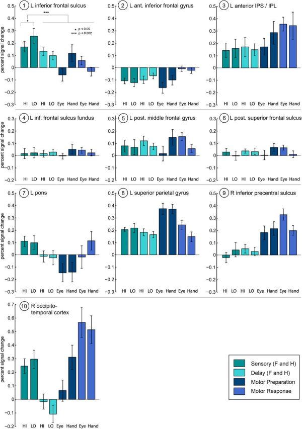
Figure 5.

Percent BOLD signal change for each area revealed by the PPI in Figure 4A. The left inferior frontal sulcus is the only region that (1) shows a modulation by the amount of sensory evidence (here: Low>High); (2) shows greater activation during the perceptual decision stage than during the rest of the trial; and (3) is positively activated during the perceptual decision stage. Other areas are deactivated (left anterior IFG), not significantly different from baseline during the decision (right inferior pre-CS, left posterior SFS, fundus of left IFS), or are not modulated by evidence levels during the sensory stage. Error bars represent the SEM (vs baseline). Note HI versus LO Sensory error bars are versus baseline, not for the difference between HI and LO. A paired two-tailed t test between HI and LO Sensory activations was significant (p < 0.05). HI, High sensory evidence (both faces and houses); LO, low sensory evidence (both faces and houses).
