Figure 7.

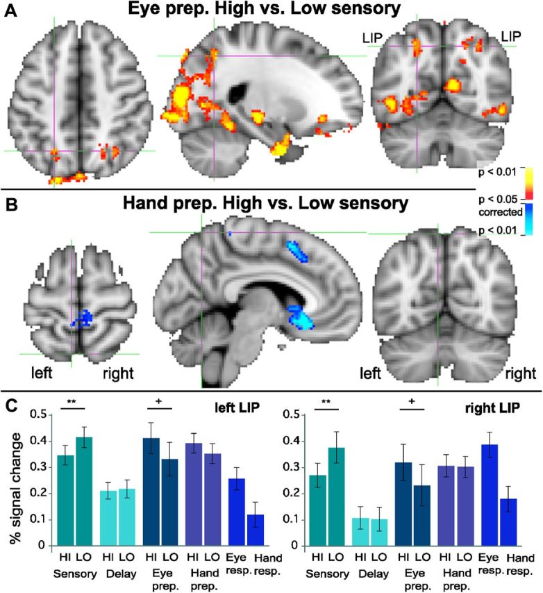
Modulation by sensory evidence during motor preparation. A, Red to yellow: putative bilateral LIP (lateral intraparietal area) shows greater activation for high sensory evidence compared with low sensory evidence, but only during the eye movement preparation stage (i.e., after the motor plan is known). B, Dark blue to light blue: areas in posterior parietal cortex, including medial parietal cortex, do not show greater activation for high compared with low sensory evidence during hand movement preparation. In contrast, motor areas such as the left pre-SMA and right paracentral lobule do show greater activation for hand movement preparation in high sensory versus low sensory trials. C, LIP shows a reversal of High>Low and Low>High modulations from the sensory stage to the eye movement preparation stage. During the decision (sensory) stage, both left and right LIP show a Low>High modulation (**paired two-tailed p = 0.008 and 0.005; t(15) = 3.03 and 3.28 for left and right LIP, respectively). In contrast, during the eye movement preparation stage, both left and right LIP show a High>Low modulation in a High>Low activation contrast (+p < 0.05, corrected at whole-brain level; within the ROI: paired two-tailed p = 0.007 and 0.009, t(15) = 3.14 and 3.02 for left and right LIP, respectively). Note that although the ROI was selected based on the latter contrast, High>Low during eye movement preparation, the contrast is significant at the whole-brain-level, corrected, before the selection of any ROI. Error bars represent the SEM.
