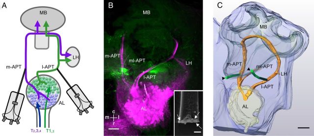
Figure 1.
Simultaneous dual-tract olfactory PN recordings using thin-wire electrodes. A, Schematic overview of the recording position in uniglomerular PN pathways of the honeybee. The AL is innervated by axonal olfactory receptor neuron tracts (T1–T4) that segregate in glomeruli of the dorsal (magenta) and ventral (green) AL hemilobes. After preprocessing in the AL, two protocerebral tracts (the m-APT and l-APT), connect glomeruli in the two hemilobes of the AL with the mushroom bodies (MB) and lateral horn (LH) in opposite order. Wire electrodes (each shaft comprising 3 copper wires, each 15 μm in diameter; see B, inset, arrows) were used to record PNs from the m-APT and l-APT. B, Staining of electrode insertion sites (green) and anterograde staining of the l-APT and m-APT (magenta). C, Reconstructions of electrode positions (either by fluorescent staining or identification of electrode tracks in the tissue) and APTs proved that on the m-APT side, the electrode was close to the m-APT, above the branching point of the mediolateral APT (ml-APT). On the l-APT side, the electrode was positioned in the lateral-caudal protocerebrum between vertical lobe and the AL (Kirschner et al., 2006, Fig. 1, positions 3 and 4). m, medial; l, lateral; c, caudal; r, rostral. Scale bars: B, C, 100 μm; B, inset, 25 μm.

