Figure 2.
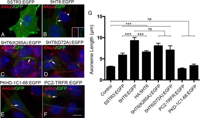
Cilia growth induced by GPCR overexpression is not significantly affected by loss of GPCR function or the presence of protein tags. (A–F) Representative confocal images of NIH3T3 cells expressing the proteins indicated above each panel whose expression was driven by the CMV promoter. All cells were immunostained with antibodies against the axoneme-enriched protein, acetylated α tubulin (AAtub) (red) and GFP (green). Nuclei were labeled with DAPI (blue). A, Cilium of a cell transfected with a vector encoding SSTR3:EGFP (arrow). B, Cilium of cell overexpressing 5HT6:EGFP (arrow) adjacent to a cilium of a nontransfected control cell (arrowhead). Insets, Single-channel EGFP and AAtub staining of the cilium of the transfected cell. C, D, Cilia elaborated by cells overexpressing the signaling defective 5HT6 receptors, 5HT6(K265A):EGFP (C) or 5HT6(D72A):EGFP (D) (arrows). E, F, Overexpression of EGFP fused to fibrocystin (PKHD-1C1–68:EGFP) (E) or human transferrin receptor (PC2-TRFR:EGFP) (F), two noncilia proteins fused to a cilia localization signal. Scale bar (in F), 10 μm. G, Mean axoneme lengths of cilia produced by cells expressing the experimental vectors shown in panels A–F and HA:5HT6. From left to right, n = 50, 34, 44, 56, 38, 22, 26, and 30 cilia analyzed/group, respectively. Error bar indicates mean ± SEM. ***p < 0.001 (one-way ANOVA). ns, Not significant.

