Figure 3.

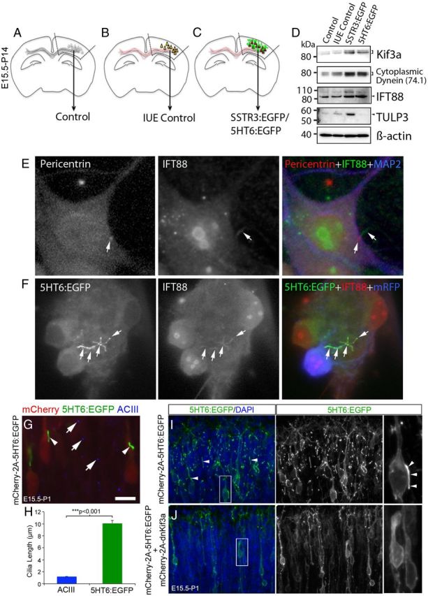
GPCR overexpression induces upregulation of IFT proteins and premature cilia lengthening. A–D, Comparisons were made between protein expression in nonelectroporated control cortex (A), fetal cortex that was electroporated at E15.5 with either a vector encoding EGFP and mCherry:AU1 (in utero electroporation control) (B), or mCherry:AU1 and either SSTR3:EGFP or 5HT6:EGFP (C). Expression of all transgenes was under the control of the EF1α promoter. Hyphenated lines indicate cortical regions of P14 brains that were used to prepare the protein lysates analyzed by Western blot. D, Western blots (10 μg of total protein/group) were probed for proteins associated with either anterograde (Kif3a) or retrograde (cytoplasmic dynein, D1 IC74) IFT complex B protein (IFT88), or GPCR trafficking into cilia (TULP3). β-Actin was used as a loading control. E, Cultured, nonelectroporated control cortical neuron immunostained for pericentrin (basal body, red), IFT88 (green), and the neuronal marker, MAP2 (blue). The arrow in the middle panel points to an IFT88+ cilium extending from a pericentrin+ basal body (arrow left panel). F, Example of an abnormally long, branched 5HT6:EGFP+ cilium synthesized by a cultured neuron expressing 5HT6:EGFP (green) under the control of the CMV promoter and mRFP (pseudocolored blue). IFT88 (red) and EGFP were colocalized along the length and branches of the cilium (white arrows). Scale bar, 5 μm. G, E15.5 brains were electroporated with a vector encoding mCherry(AU1)-2a-5HT6:EGFP. At P1, electroporated brains were sectioned and stained with an antibody against ACIII. Examination of the upper layers of the cortical plate revealed mCherry+ neurons (red) that possessed longer 5HT6:EGFP+ cilia (arrowheads) than their neighboring nonelectroporated cells whose ACIII-stained cilia appear punctate (blue, arrows). Scale bar, 10 μm. H, Comparison of the lengths of the cilia of neurons overexpressing 5HT6:EGFP and control neurons: ***Student's t test. (I) Section of brain electroporated and processed as described for G, but not including the red channel used to visualize mCherry. Numerous, often long cilia (arrows) were present in the upper layers of the cortical plate. J, P1 neurons in the upper cortical plate that were coelectroporated at E15.5 with vectors encoding mCherry and 5HT6:EGFP (mCherry(AU1)-2a-5HT6:EGFP) and mCherry and dnKif3a (mCherry(AU1)-2a-dnKif3a). The elongated 5HT6:EGFP+ cilia of neurons expressing 5HT6:EGFP alone (I) are not observed in cells coexpressing 5HT6:EGFP and dnKif3a.
