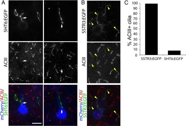
Figure 5.
Cilia of neurons overexpressing 5HT6:EGFP do not contain detectable levels of ACIII. The brains of E15.5 embryos were electroporated with vectors encoding either 5HT6:EGFP or SSTR3:EGFP under control of the EF1α promoter and were immunostained for ACIII at P14. A, Pyramidal neurons in layers 2/3 of neocortex expressing mCherry:AU1 (blue) and 5HT6:EGFP (green). Sections were immunostained for ACIII (red), which normally is enriched in cilia of virtually all neocortical neurons. White arrows point to 5HT6:EGFP+ cilia projecting from mCherry:AU1+ neurons that lack detectable ACIII staining. Scale bar, 10 μm. B, Pyramidal neurons in layers 2/3 of neocortex expressing mCherry:AU1 (blue) and SSTR3:EGFP (green). SSTR3:EGFP+ cilia also stain for ACIII (yellow arrowheads). C, The percentage of SSTR3:EGFP+ (n = 123) or 5HT6:EGFP+ (n = 89) cilia that are also ACIII+.

