Figure 7.

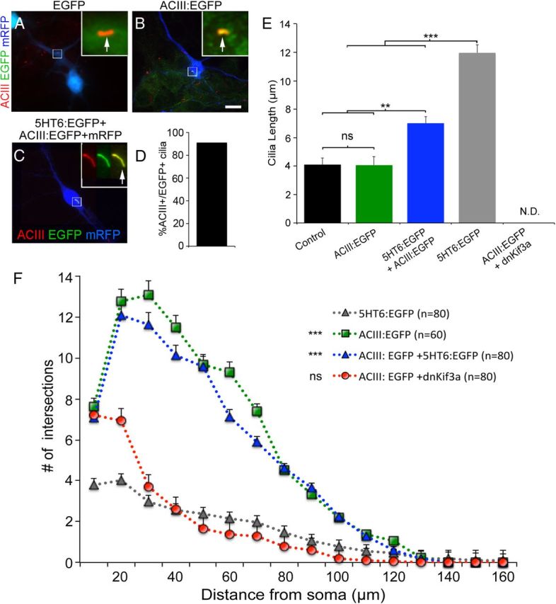
Coexpression of ACIII:EGFP with 5HT6:EGFP, but not dnKif3a, restores ciliary ACIII, cilia structure, and dendrite outgrowth. A, B, Neurons electroporated at E15.5 with vectors encoding either (A) mRFP and EGFP or (B) mRFP and ACIII:EGFP. Electroporated neurons were cultured for 6 DIV, fixed, and immunostained for ACIII (red). Analyses of mRFP+ neurons (blue) showed that EGFP did not traffic into the cilia as evidenced by an absence of colocalization with ACIII (A; arrow in inset). When fused to ACIII, the cilia were positive for both ACIII staining and EGFP fluorescence (B; arrow in inset). Scale bar (in B), 10 μm. C, Example of a cultured neuron coelectroporated with 5HT6:EGFP and ACIII:EGFP possessing a cilium that is positive for both ACIII staining and EGFP (inset shows higher magnification of cilia in individual channels and merge). D, Quantification of the number of cells coelectroporated with 5HT6:EGFP and ACIII:EGFP whose cilia were both ACIII+ and EGFP+ (N = 139 of 153). E, Comparisons of the lengths of the cilia elaborated by neighboring nonelectroporated (control) neurons (n = 142) or neurons transfected with vectors encoding mRFP plus ACIII:EGFP (n = 60), ACIII:EGFP and 5HT6:EGFP (n = 120), 5HT6:EGFP (n = 120), or dnKif3a (n = 118) and cultured for 12 DIV. **p < 0.01. ***p < 0.001. ns, Not significant; N.D., not determinable. F, Sholl analyses of the complexity of the dendritic arbors elaborated by neurons transfected with mRFP plus either 5HT6:EGFP (gray), ACIII:EGFP (green), EGFP:dnKif3a + ACIII:EGFP (red), or ACIII:EGFP + 5HT6:EGFP (blue) and maintained in culture for 12 d (n = number of cells analyzed). The complexity of the arbors of neurons expressing 5HT6:EGFP were statistically compared with those of the other groups using two-way ANOVA: ***p < 0.001. ns, Not significant).
