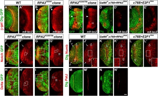
Figure 9.
Notch signaling activity is downregulated in RPA and E2F1 mutant NE cells. Brain lobes from late-third instar larvae were stained with the antigens indicated; the clones are marked by the lack of GFP expression and by dashed lines. Arrow indicates the lamina furrow (LF). A, A', E(spl)m8-lacZ expression in wild type. B–C', E(spl)m8-lacZ expression was dramatically downregulated in RPA3 (B, B') or RPA2 (C, C') mosaic NE clones (filled arrowheads). There is also a medulla clone immediately underneath the NE that had lost E(spl)m8-lacZ expression (C', open arrowhead). D–E', E(spl)m8-lacZ expression in RPA3RNAi brains (D, D') and E2F1RNAi brains (E, E'). E(spl)m8-lacZ expression was downregulated in RPA3 or E2F1 mutant brains. F–G', Notch expression was not affected in RPA3 (F, F') or RPA2 (G, G') mutant epithelial clones. H–I', Delta expression was not influenced in RPA3 (H, H') or RPA2 (I, I') mutant epithelial clones. Note that in mutant clones located entirely in the medulla, there was an increase of both Notch and Delta expression (G', I', double arrowheads). J–L', Numb localization in wild type (J, J'; yellow arrows indicate the apical membrane domain of NE cells), RPA3RNAi brains (K, K'), and E2F1RNAi brains (L, L'). Open arrows indicate the basal domain of NE cells. Numb is mislocalized and strongly concentrated along the subapical region and the lateral region of the NE cells in RPA3 and E2F1 mutant brains. M–N′, PatJ expression in wild-type (M, M') and RPA3RNAi brains (N, N′). The apical marker PatJ was reduced or almost eliminated in RPA3RNAi brains (N, N′). Lateral is to the left and dorsal up. Scale bar, 20 μm.

