Figure 2.

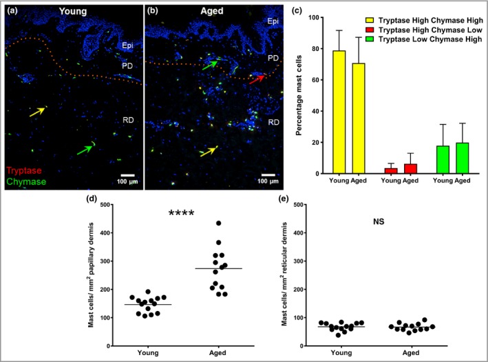
Increased mast cell (MC) numbers in aged skin are localized to the papillary dermis but show no change in protease phenotype. Dual immunohistochemistry for MC tryptase (red) and MC chymase (green) in (a) young and (b) aged skin. Some MCs show strong immunofluorescence for both enzymes (yellow arrows) while others are chymase (green arrows) or tryptase dominant (red arrow). (c) The proportions of MC phenotypes were the same for both age groups (mean ± SEM percentage); however, an age‐related increase in numbers was found in the papillary dermis (d) but not the reticular dermis (e). Epi, epidermis; PD, papillary dermis; RD, reticular dermis. Data from 14 young and 13 aged individuals; ****P < 0·001; NS, not statistically significant using the unpaired two‐tailed Mann–Whitney U‐test.
