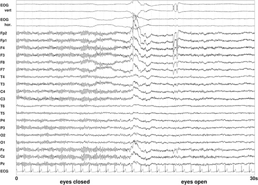
Figure 7.

Resting‐state electroencephalography (EEG) with eyes closed (left) and eyes open (right). Figure shows a 30‐s epoch with 19 EEG signals referenced to the averaged mastoids, a bipolar vertical and horizontal electrooculogram (EOG), and an electrocardiogram (ECG). The blockade of alpha in the EEG by opening of the eyes is obvious.
