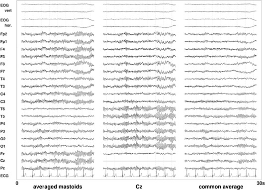
Figure 9.

Examples of the impact of different references on the resting‐state electroencephalography (EEG). In the averaged mastoids montage, for most locations comparably high alpha amplitudes occur, with the lowest amplitudes for the temporal and frontal regions. With the Cz (single electrode) reference, the central and frontal regions show lower alpha amplitudes (the Cz location itself is zero in this diagram, of course.) A common average reference (mean of all scalp locations) leads to a generally weaker alpha rhythm.
