Figure 3.
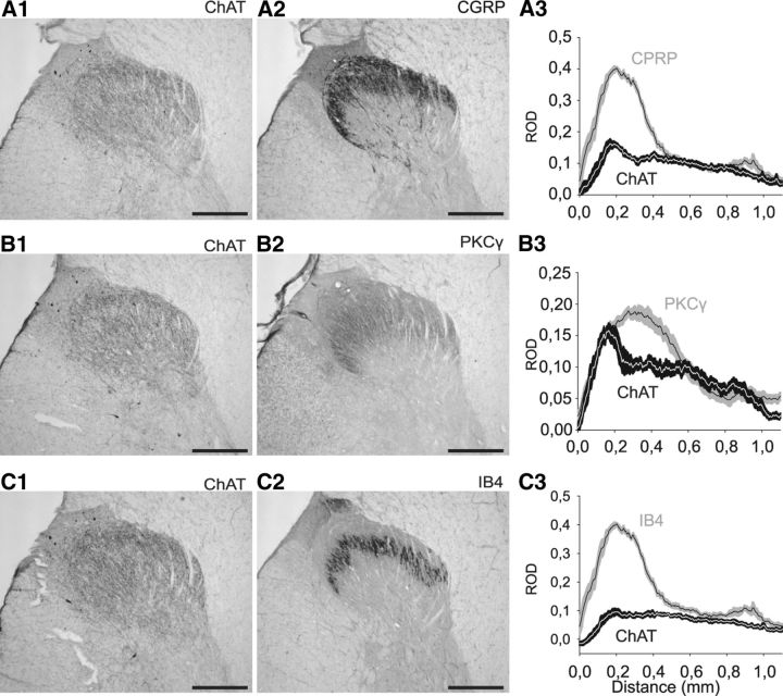
ChAT-immunopositive plexus compared with anti-CGRP, anti-PKCγ, or anti-IB4 immunolabeling in monkey lumbar spinal dorsal horn. A, Comparison of anti-ChAT (A1) and anti-CGRP immunolabeling (A2) on adjacent transverse sections of the dorsal horn (30-μm-thick). Graphic representation (A3) of the mean ± SEM ROD of ChAT (black) and CGRP (gray) staining (six sections, one animal). B, Comparison of anti-ChAT (B1) and anti-PKCγ (B2) immunolabeling on adjacent transverse sections of the dorsal horn (30-μm-thick). Graphic representation (B3) of the mean ± SEM ROD of the ChAT (black) and PKCγ (gray) stainings (six sections, one animal). C, Comparison of anti-ChAT immunolabeling (C1) and IB4 staining (C2) on adjacent transverse sections of the dorsal horn (30-μm-thick). Graphic representation (C3) of the mean ± SEM ROD of ChAT (black) and IB4 (gray) staining (six sections, one animal). Nonspecific deposits of DAB reaction product are observed in the upper left edge of lamina I (next to the dorsal root entry zone) in A1, B1, and C1. Scale bar, 500 μm.

