Figure 8.

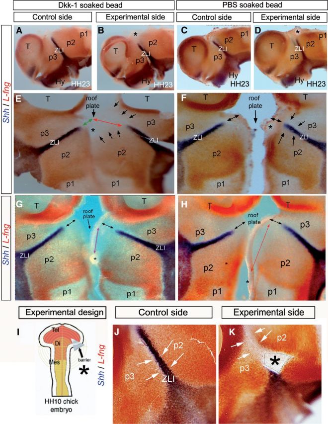
Wnt signaling is required for L-fng encroachment in the alar diencephalon. A–D, J, K, Lateral views of the whole-mount prosencephalic neural tube stained for Shh (blue) and L-fng (red) by double ISH at HH23. A, C, J, Control side of the experimental embryos. B, D, K, Experimental side of the embryos. E–H, Flat mounts opened through the ventral midline. Asterisk in B, E, and G indicates the region in which a Dkk-1-soaked bead was implanted at HH10. Asterisk in D, F, and H indicates the region in which the PBS-soaked bead was implanted. Green and red arrows in E indicate the distance between the dorsal midline and the dorsal level of Shh expression in the control (green) and experimental (red) side of an embryo in which a Dkk-soaked bead was implanted (asterisks). Black arrows in E indicate that L-fng encroachment was disrupted in the experimental side of the embryo. Black arrows in F and H indicate the similar distance between the dorsal midline (roof plate) and the dorsal level of Shh expression in the control and experimental side of an embryo in which the PBS-soaked bead was implanted. Black arrows in G indicate the same distance between the dorsal midline (roof plate) and the dorsal level of Shh expression in the control and experimental side of an embryo in which the Dkk-1-soaked bead was implanted in the most caudal position of the alar/roof plate of p2. I, Schematic representation of barrier implantation in HH10 chick embryo. J, Lateral view of the control side of HH23 chick embryos analyzed by double ISH for Shh (blue) and L-fng (red). K, Lateral view of the experimental side of chick embryos. Asterisk in K indicates the region in which the microbarrier was implanted at HH10. Arrows in J and K indicate that the expression of L-fng at both sides of the ZLI was not disrupted in embryos operated with the microbarrier, in which Shh was blocked to progress through the ZLI. Di, Diencephalon; Hy, hypothalamus; Mes, mesencephalon; T, Tel, telencephalon.
