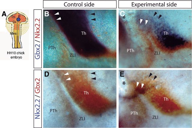
Figure 9.
Inhibition of Wnt signal results in downregulation and ectopic expression of Gbx2 and Nkx2.2. A, Schematic representation of the neural tube explants in which Dkk-1 was inserted. B–E, Lateral view of the control side (B, D) and experimental side (C, E) of chick embryos neural tube analyzed by ISH for Gbx2 in blue and Nkx2.2 in red (B, C) and for Nkx2.2 in blue and Gbx2 in red (D, E). Black arrowheads localize the caudal limit of Gbx2 expression (see its reduction in C and E), whereas white arrowheads localize the region in the dorsal ZLI in which Gbx2 and Nkx2.2 were ectopically expressed. Black asterisks indicate the place in which Dkk-1-soaked beads were implanted. Th, Thalamus; PTh, prethalamus.

