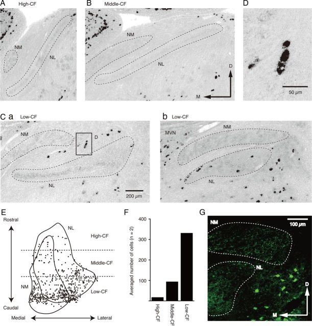
Figure 3.
GABAergic neurons are clustered in the low-CF region of the NL. A–C, Distributions of GAD1 mRNA-positive cells detected by in situ hybridization. The slices contain high-CF (A), middle-CF (B), and low-CF (C) regions of the NL. The broken lines indicate areas of the NM and NL. D, High magnification of the box in Ca. E, Two-dimensional distribution of GAD1 mRNA-positive cells in and around the NM and NL. F, The average number of GAD1 mRNA-positive cells around each CF region of the NL (separated by broken lines in E). G, Immunoreactivity to the GABA antibody in the lateral parts of the NM and NL regions. The rostrocaudal level of the slice is the same as that in Cb.

