Figure 5.

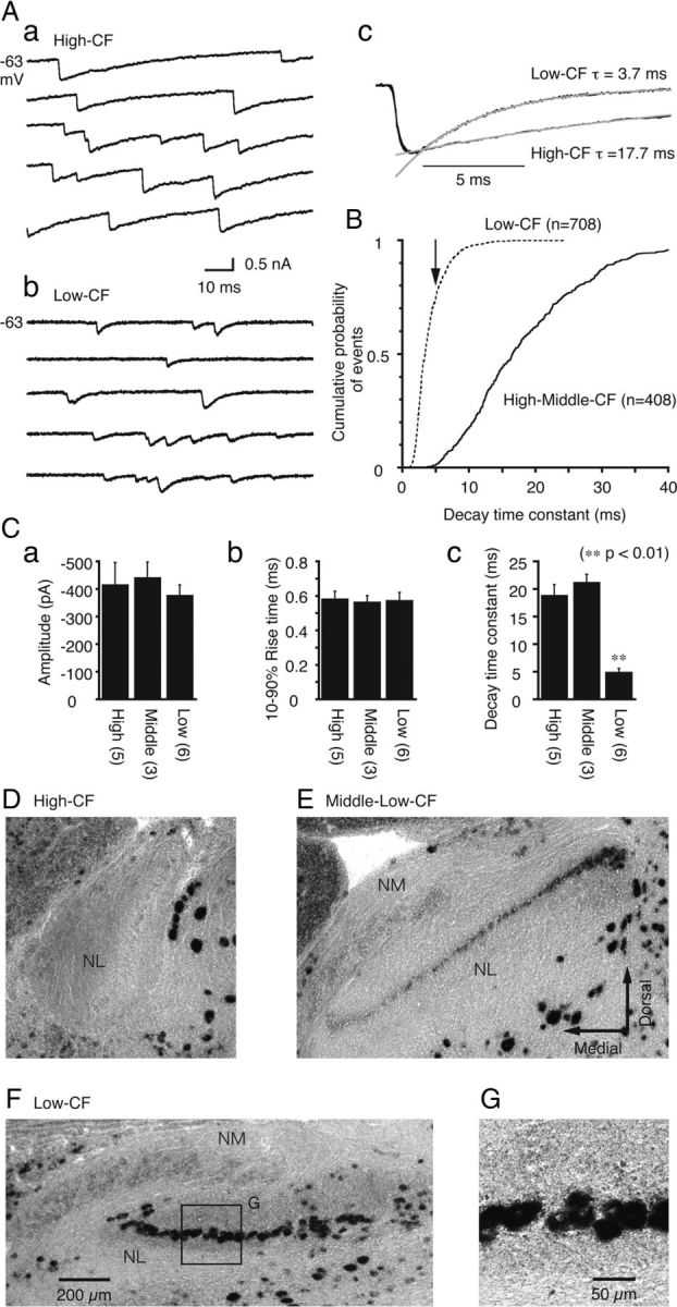
Fast decay kinetics of sIPSCs and strong expression of the GABAA receptor α1-subunit in low-CF neurons. A, Representative traces of sIPSCs recorded from a high-CF (a) and a low-CF (b) neuron. sIPSCs from these two neurons were extracted, ensemble-averaged, and superimposed after normalization (c). The gray lines are the single-exponential fit to the decay time course. B, Cumulative probability plots of the sIPSC decay time constants sampled from high-CF and middle-CF (solid line; 408 events from 8 cells) and low-CF (broken line; 708 events from 6 cells) neurons. The arrow indicates the averaged time constant of the low-CF neurons. C, Averaged sIPSC values including the amplitude (a), 10–90% rise time (b), and decay time constant (c), were compared among the high-CF, middle-CF, and low-CF neurons. The decay time constant of the low-CF neurons was significantly different from the other CF neurons (p < 0.01). D–F, The expression patterns of the GABAA receptor α1-subunit mRNA as detected by in situ hybridization. Representative coronal slices that comprise the high-CF (D), middle-low-CF (E), and low-CF (F) regions of the NL. The lower CF regions in the NL were close to the lateral edge (Rubel and Parks, 1975). G, High magnification of the box in F.
