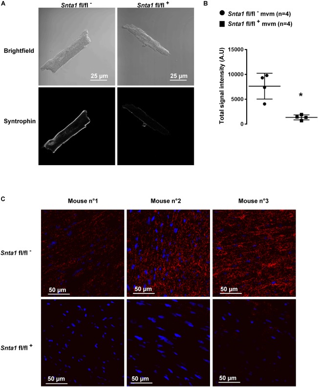
FIGURE 2.
α1-Syntrophin staining in wild-type and knockdown α1-syntrophin knockdown cardiac sections and cardiomyocytes. (A) Immunostaining, using a pan-syntrophin antibody, of syntrophin in wild-type (Snta1 fl/fl-) and cardiac-specific α1-syntrophin knockdown murine cardiomyocytes (mvm) (Snta1 fl/fl+). (B) Intensity quantification of syntrophin signal of four cells confirming the significant decrease of syntrophin in knockdown mice. (∗p < 0.05). The number of cells is indicated in parentheses. (C) Duolink® experiments showing a significant decrease in interaction (red dots) between Nav1.5 and syntrophin in α1-syntrophin cardiac specific knockdown cardiomyocytes (Snta1 fl/fl+) compared to wild-type (Snta1 fl/fl-).

