Figure 3.

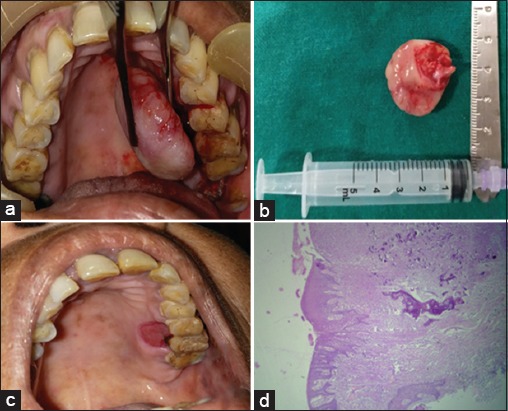
(a) Intraoperative photograph, (b) Excised specimen, (c) Post-operative site of lesion, (d) Photomicrograph of lesion showing epithelium overlying connective tissue stroma comprising of bony trabeculae with osteoblastic rimming (×4)

(a) Intraoperative photograph, (b) Excised specimen, (c) Post-operative site of lesion, (d) Photomicrograph of lesion showing epithelium overlying connective tissue stroma comprising of bony trabeculae with osteoblastic rimming (×4)