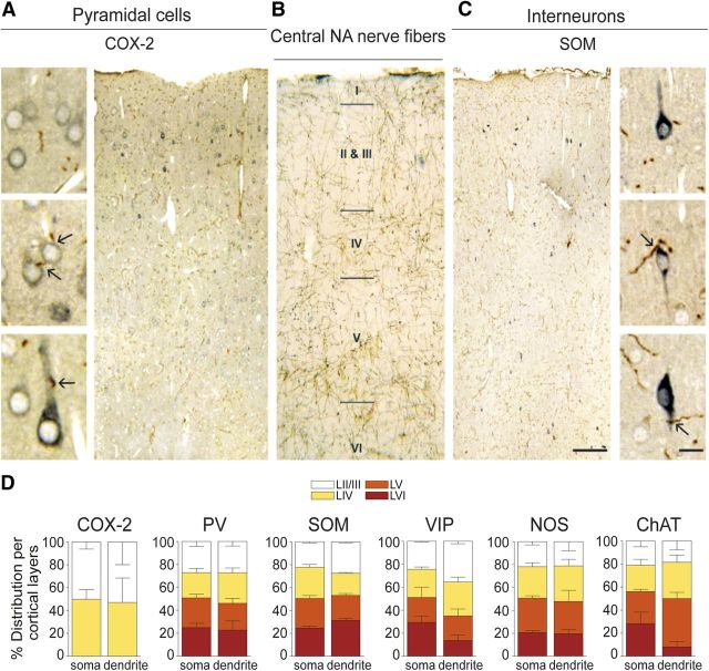
Figure 1.
NA innervation of cortical COX-2 pyramidal cells and distinct subsets of GABA interneurons. A, C, Photomicrographs of semithin, double-immunostained sections for NA fibers and COX-2 pyramidal cells or SOM interneurons. The brown DAB-immunodetected NA fibers contact the blue–gray SG-stained neurons on their cell soma or proximal dendrites (black arrows on high-magnification pictures). B, Photomicrographs of DBH-immunostained NA innervation in the frontoparietal cortex detected with DAB. D, Quantitative analysis of NA fibers contacting cortical COX-2 pyramidal cells and PV-, SOM-, VIP-, NOS-, or ChAT-expressing GABA interneurons. Cells were counted across all cortical layers except for COX-2 pyramidal cells that distribute mainly in layers II/III and IV. The percentage of cells innervated on their cell soma or proximal dendrites was comparable across all cortical layers. Values are mean ± SEM. Scale bars: 100 μm; high magnification, 10 μm.

