Figure 3.

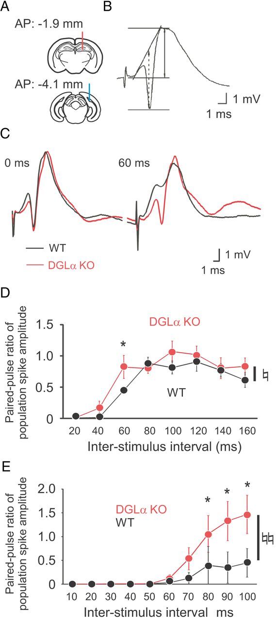
Reduced paired-pulse depression of population spike in freely moving and anesthetized DGLα knock-out (KO) mice. A, Schematics diagram showing the positions of recording (top, red line) and stimulation electrodes (bottom, blue line). B, An example of the dentate field potentials evoked by medial perforant path stimulation and recorded from one position in the dentate hilus of a wild-type (WT) mouse. The two main components of this potential are the initial positive deflection of the fEPSP and the negative-going population spike. Amplitude of fEPSP is indicated as arrows with solid line, and that of population spike is indicated as arrows with dotted line. C, Sample traces of the dentate field potentials evoked by paired-pulse stimuli with interpulse interval of 60 ms in a DGLα KO and a WT mouse. D, Ratio of population spike amplitude (second spike amplitude/first spike amplitude) evoked by paired-pulse stimuli with interstimulus intervals of 20–160 ms in DGLα KO (n = 8) and WT (n = 8) mice. E, Ratio of population spike amplitude evoked by paired-pulse stimuli with interstimulus intervals of 10–100 ms in DGLα KO (n = 5) and WT (n = 5) mice under isoflurane anesthesia. Error bars indicate means ± SEM. ♮p < 0.05 and ♮♮p < 0.01 for the effect of genotype in two-way ANOVA. *p < 0.05 in Tukey's test.
