
Figure 7.
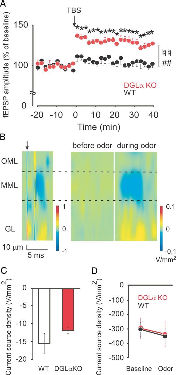
Enhanced LTP but unchanged odor-induced synaptic inputs to the dentate gyrus in DGLα knock-out (KO) under urethane anesthesia. A, Time course of fEPSP amplitude in the dentate gyrus in DGLα KO (n = 5) and wild-type (WT) (n = 6) mice before and after TBS (arrow). B, Color maps showing CSD in the dentate gyrus after perforant path stimuli (left), and before (middle) and during (right) exposure to odor. Outer molecular layer (OML), middle molecular layer (MML), and granule cell layer (GL) are separated by dotted lines. The arrow on the left indicates the perforant path stimulation. Current sink (blue) after perforant path stimulation corresponds to inward membrane current of the MML. C, Total sink current of MML and OML during 10 ms after perforant path stimulation in DGLα KO (n = 6) and WT (n = 6) mice. D, Total sink current of MML and OML during 10 s before and during odor stimulation in DGLα KO (n = 6) and WT (n = 6) mice. Error bars indicate means ± SEM. ♮♮p < 0.01 for the effect of genotype, ##p < 0.01 for the interaction between genotype and time in two-way ANOVA, and *p < 0.05 in Tukey's test.