
Fig 4. Choanomastigote forms observed in axenic culture of the LBT 7439 sample.
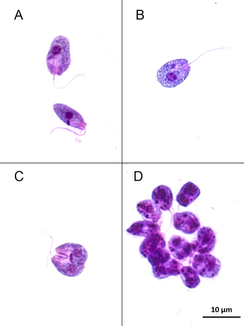
(A) Parasite cells show a typical collar-like extension through which a single flagellum emerges. (B) The kinetoplast is anterior to the nucleus and adjacent to the flagellar pocket where emergence of the flagellum can be observed. (C) Dividing forms. (D) Rosette of flagella.