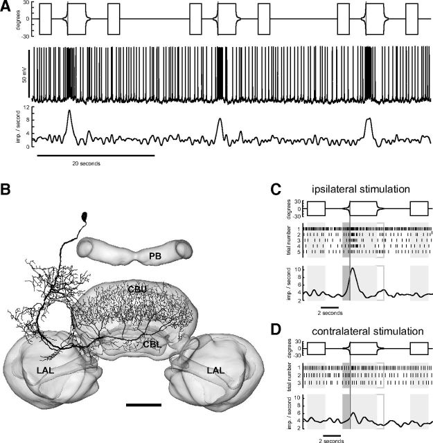
Figure 3.
Looming-sensitive TUy tangential neuron of the CBU. A, Recording trace (middle) during stimulation of the ipsilateral eye. Lower trace shows Gaussian-filtered spike train. Upper trace shows angular extent of disc as a function of time. B, Frontal reconstruction of the TUy neuron, registered into the standard central complex of the locust brain (el Jundi et al., 2010). C, D, Mean Gaussian-filtered spike trains (lower traces), corresponding raster plots (middle), and stimulus regimes (upper traces) for ipsilateral (C) and contralateral eye stimulation (D). Shaded and framed boxes and vertical lines as in Figure 1. The three trials shown in A correspond to trials 2, 3, and 4 shown in C. LAL, lateral accessory lobe. Scale bar, 100 μm.

